
Why this matters
Making viable eggs from stem cells has already been accomplished in mice. In 2016, our collaborator Katsuhiko Hayashi demonstrated that mouse skin cells can be turned into ‘induced pluripotent stem cells’ (iPSCs, which are engineered cells capable of becoming any kind of cell in the body) and then turned into usable eggs. These eggs produced healthy pups that lived normal lifespans and reproduced naturally, having healthy pups of their own.

Figure 2 – Adult mice from eggs derived from pluripotent stem cells (Hikabe et al., 2016)
This process, known as "in vitro gametogenesis” (IVG), has been far easier to achieve in mice than in larger animals. Still, given how dramatically impactful this technology could be, it is well worth pursuing for human application.
IVG has the potential to redefine reproduction worldwide. From a simple blood draw, one could make as many healthy eggs as a family needs.
This capability could create freedom from biological and genetic limits. It could dramatically expand families’ options for having healthy children and enable women to have children at a much older age– all without the hormone injections or surgical retrieval currently required for IVF.
The technology is one of the most complex therapies ever to be developed. We are not making just a single cell type; we are building entire mini-ovaries in the lab derived from stem cells, as the whole organ is important for proper egg development. We’re excited that we’ve made hugely significant progress towards this goal, and we wanted to share a peek into our process.
Our Approach: Making mini-ovaries in the lab

Figure 3 – Conception's overall process for making egg cells from stem cells.
Conception's thesis is simple: there are no useful shortcuts. A cell that expresses a few egg markers is not enough. We need to rebuild, as closely as possible, the sequence that nature uses — and benchmark our cells against human development at every major step.
Our approach follows the major steps of egg development above in Figure 3. After taking a blood sample, we turn a subset of blood cells into iPSCs, and then guide the iPSCs toward becoming each of the kinds of cells found in a developing ovary: ‘primordial germ cells’ are the cells that will eventually become eggs, and ‘ovarian helper cells’ are the supporting players that provide essential signals for the eggs. Together, these cells form ‘mini-ovaries,’ small 3-dimensional “balls of cells” that mimic a true human ovary.
Below on the left, you can see what our mini-ovaries look like with the naked eye. The middle image shows thin slices of those same mini-ovaries on a microscope slide; each white circle is one slice of a mini-ovary. These slides are then used for our image analysis on the right where we stain the mini-ovaries with cell and stage-specific dyes to understand how they are developing.
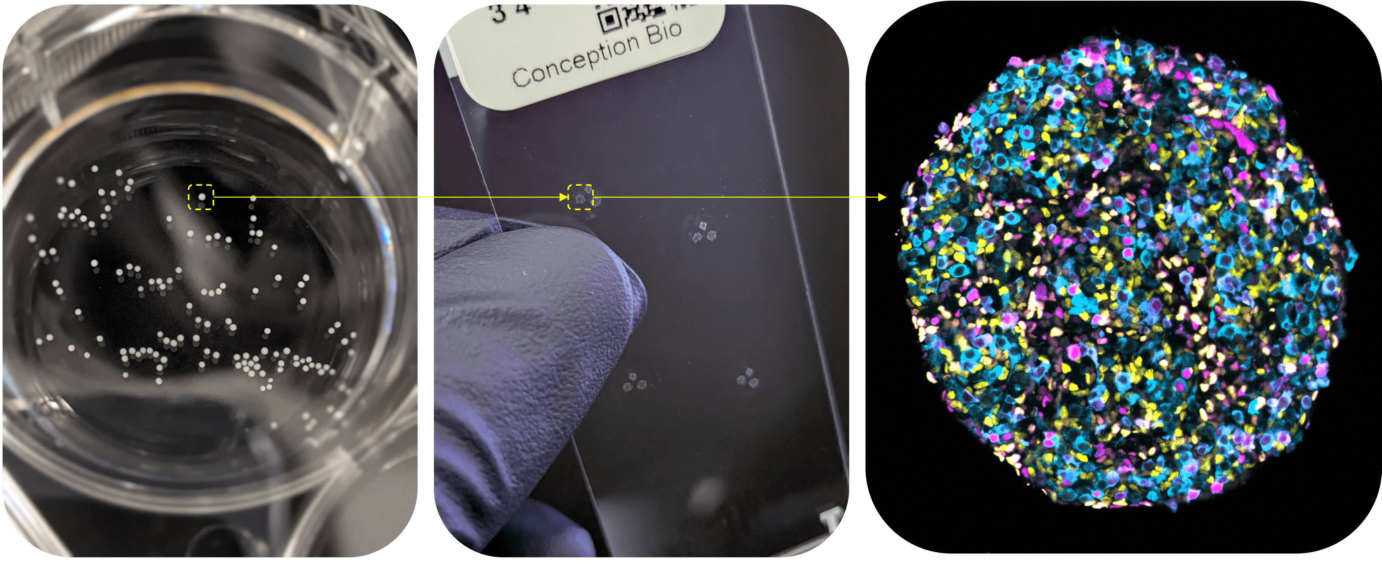
Figure 4 – Examples of Conception's mini-ovaries
In our research, we generate thousands of mini-ovaries, containing millions of future egg cells, to study, improve, and benchmark their development in parallel.
Inside the mini-ovaries, primordial germ cells are surrounded by the ovarian helper cells they need to begin moving through the next three stages of egg development:
1) The primordial germ cells progress toward ‘oogonia’
2) The oogonia enter into meiosis, the special cell division needed to make eggs
3) As they become early egg cells, they form follicles, the essential ovarian units that house each egg
Along the way, we rigorously benchmark cell identity against a massive internally-assembled reference atlas of human ovary molecular data. This atlas includes millions of datapoints spanning a wealth of molecular features capturing many layers of cell biology, from which genes are turned on to the epigenetic features that regulate cell identity. Comparisons to this atlas (including with proprietary deep learning models) allow us to confidently chart our path forward biologically, while confirming the fidelity of our protocol and thus the quality of our cells.
One of the most important measures of success for us is function - can these cells faithfully perform the same roles of cells in a real ovary? We’ll walk through how we benchmark that in each step below.
1) Our mini-ovaries help develop oogonia
An early sign of success for our mini-ovaries is that we see their organization closely mimics the structure of a developing human ovary. Oogonia form small “nests” – special ovarian structures surrounded by a thin boundary layer (in blue below) where future egg cells stay connected in groups and chains (in magenta). In the ovary, these structures help separate and organize developing egg cells, so seeing them form in our mini-ovaries is a sign that the tissue is developing the same way as it would in the human body.

Figure 5 - Example of Conception's stem cell-derived mini-ovary (left) compared to natural human ovary (right).
All of the cells shown on the left were derived from stem cells. They independently start forming these ovarian structures without any natural human cells in the culture, and without forcing the cells artificially into these shapes. We find this remarkable to observe.
2) Our future egg cells progress through meiosis
Most cells in our body contain two sets of chromosomes - one inherited from each parent - whereas egg cells contain only one. Meiosis is one of the defining events in egg development, and it’s how the egg ends up with one set of chromosomes. It must happen with extraordinary precision because chromosomal mistakes can lead to failed pregnancies or genetic abnormalities.
Meiosis is one of the hardest things to get right. Chromosomes have to pair with their matching partners, exchange DNA, and (in the body) remain organized for decades. This is why the next result was so important to us: in our iPSC-derived cells, we see the machinery of meiosis assembling as it should.

Figure 6 - Meiosis I progression steps. Conception's stem cell-derived (top and right) vs. natural human (bottom) future egg cells.
Stem cell-derived germ cells show assembly of the meiotic chromosome-pairing machinery. This is an essential proof point for any credible path toward human IVG.
A useful way to picture this process is as a zipper forming along each chromosome pair. In our cells, key structural proteins of the meiotic machinery load onto chromosomes in long, continuous tracks, consistent with the cells progressing through early meiosis.
This is not just a gene marker turning on. It is cellular machinery appearing in the right place, in the right biological context, inside a fully stem cell-derived ovarian system.
We also see the broader molecular signatures expected as our cells transition toward early egg cells. We see key primary oocyte genes activate, including genes involved in egg growth, communication with helper cells, formation of the zona pellucida (the protective “egg shell” around the oocyte), and programs that help protect developing eggs.

Figure 7 - Early-egg cell markers. Conception's stem cell-derived (top) vs. natural human (bottom) early-egg cells in follicles.
Together, this all shows that our stem cell-derived cells are moving through meiosis and activating early egg cell genes as should be properly happening at this stage.
3) We can make fully iPSC-derived follicles
After entering meiosis, future eggs in the human ovary enter a long resting period. At this stage, the cell helps form a primordial follicle: one egg cell surrounded by a single layer of tightly connected support cells. This is the basic and most important unit of the ovary.
Below you can see on the left, side by side with follicles from an actual human ovary on the right, what we believe are the first human follicles ever created entirely from iPSCs. The developing egg cells are shown in magenta, the surrounding support cells are shown in yellow, and the blue shows the thin boundary that wraps around each follicle. As the early egg cells undergo meiosis, the yellow support cells attach to them and begin to nurture them. They organize into a single flattened layer around each early egg cell and deposit the thin boundary, recreating the defining structure of early human ovarian development.

Figure 8 - Early-egg cell markers - Conception's stem cell-derived follicles (left) compared to natural human follicles (right).
Generating fully stem cell-derived follicles, with early egg cells progressing through meiosis, is a major step toward making viable mature eggs. To our knowledge, this is a world first.
What’s next for stem cell-derived eggs
While we’ve come a long way, there is still more work to be done. The biggest remaining step for us is to grow our iPSC-derived follicles from the early stage (primordial) to the last “antral” step. At the antral stage, the oocytes have grown larger and are at the point where an IVF physician would collect them surgically. We believe this should be quite doable, as we have previously accomplished this with donated human tissue (below).
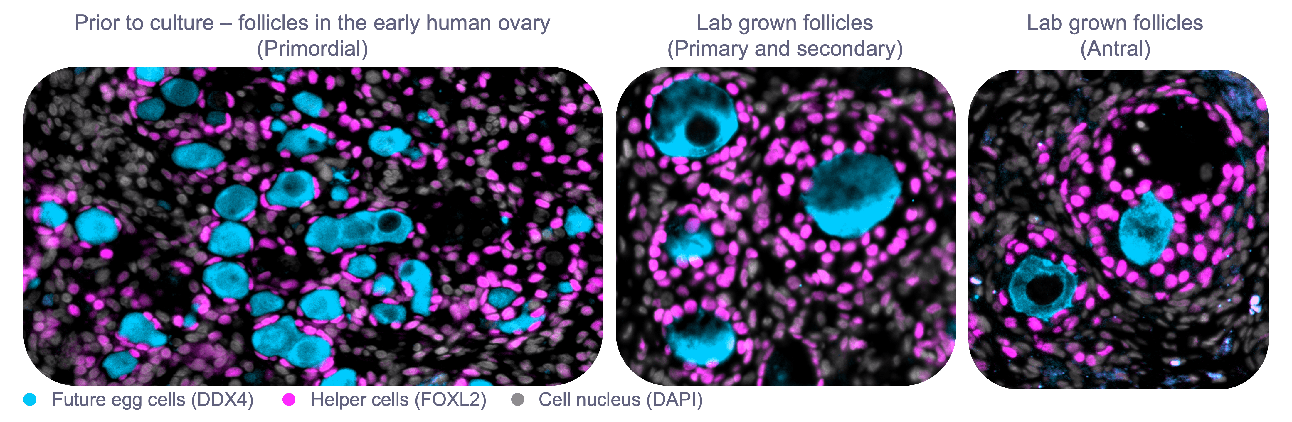
Figure 9 - Lab grown human follicles from early stage developing human ovary. Primordial to antral follicle stages.
Beyond that, our focus will be on validating the safety of our process and quality of our eggs. The bar for safety with this technology is incredibly high, and we take that responsibility very seriously. Before this work could be considered for clinical use, we need to deeply characterize each step of the process, both for existing progress and for fully mature egg cells in the future. This includes deeper animal model development and validation for safety as well.
If you think this is cool, please reach out
We are very excited to share a small taste of what we’re working on, and we would love to hear from you if you could benefit from our work. Feel free to email us at hello@conception.bio.
And if you think you have the skills to contribute, please take a look at our job openings. We believe this is the most challenging and exciting research project in biotech, and it could end up as one of the most impactful technologies of our lifetimes. We are very actively hiring, so if a role looks like it could be a fit, please apply or email us.